Orbital Decompression

What Is Orbital Decompression Surgery?

Orbital decompression surgery, also known as decompression surgery, is a surgical procedure performed to primarily address proptosis or bulging eyes. It is commonly performed for symptoms and complications associated with Thyroid Eye Disease (TED), also known as Graves’ ophthalmopathy or Graves’ orbitopathy, an autoimmune disorder in which the tissues around the eyes become inflamed and enlarged, causing a range of symptoms, including proptosis, double vision, and eye discomfort. The procedure involves removing some of the bony structures or fat within the eye socket to create additional space for the swollen eye muscles and fatty tissues, allowing the eyes to move back into a more normal position and relieving pressure on the optic nerve. This can alleviate symptoms and lead to improved eye appearance and function.

What Are the Different Types of Orbital Decompression?

There are several different types of orbital decompression surgery, each with its approach and specific goals. The choice of which type of surgery to perform depends on the individual patient’s needs, the severity of the condition, and the surgeon’s expertise. The main types of orbital decompression surgery include:

  • Orbital Fat Decompression, also known as fat decompression or orbital fat removal, is a surgical procedure that removes excess fatty tissue from the eye socket (orbit) to alleviate symptoms and improve the appearance of the eyes in individuals with Thyroid Eye Disease (TED) or other conditions associated with bulging eyes.
  • Orbital Bone Decompression involves removing some of the bony structures within the eye socket (orbit) to create additional space for the swollen eye muscles and fatty tissues, allowing the eyes to move back into a more normal position and relieving pressure on the optic nerve.

When Is Orbital Decompression Surgery Indicated?

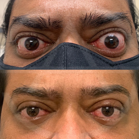
Orbital decompression surgery is primarily performed for medical reasons to address eye-related symptoms and complications associated with conditions like Thyroid Eye Disease (TED) or other disorders that cause proptosis (bulging eyes). The primary goals of orbital decompression surgery in these cases are to:

  • Relieve pressure on the optic nerve: In severe cases, the bulging eye tissues can compress the optic nerve, leading to vision problems. Orbital decompression can alleviate this pressure, protecting the optic nerve and preserving vision.
  • Improve eye alignment and function: Bulging eyes can cause double vision, difficulty closing the eyes, and other functional issues. By creating additional space within the eye socket, orbital decompression can help improve eye alignment and functionality.
  • Reduce eye discomfort and symptoms: People with bulging eyes often experience eye pain, redness, swelling, and dryness. Orbital decompression can help alleviate these symptoms and improve overall eye comfort.

While orbital decompression surgery is primarily medical in nature, it can have a positive impact on the cosmetic appearance of the eyes. By reducing proptosis and restoring a more natural eye position, the procedure often results in a more pleasing and balanced facial appearance.

Orbital Decompression Procedure Overview:

Orbital decompression surgery is primarily used to address bulging eyes or proptosis. The procedure involves removing some of the bony structures within the eye socket (orbit) to create additional space for the swollen eye muscles and fatty tissues, allowing the eyes to move back into a more normal position and relieving pressure on the optic nerve. This can lead to improved eye appearance and function and alleviate symptoms.

Here’s an overview of the procedure:

  • Patient Evaluation: Before undergoing orbital decompression surgery, patients undergo a thorough evaluation by an ophthalmologist and an oculoplastic surgeon. This evaluation includes measuring the degree of proptosis, assessing eye movement, and considering the patient’s overall eye health and condition.
  • Surgical Approach: The surgery is done under general anesthesia and can be performed through various approaches, including through the upper eyelid (transpalpebral), the lower eyelid (transconjunctival), or a combination of both. The choice of approach depends on the patient’s specific condition and the surgeon’s preference.
  • Recovery Bone Removal: During the surgery, the surgeon removes a portion of the bony walls of the eye socket. Typically, the medial (inner) wall and/or the lateral (outer) wall of the orbit are addressed. By removing these sections of bone, the orbital volume is increased, creating space for the swollen tissues.
  • Eye Realignment: The removal of bone allows the enlarged eye muscles and fatty tissues to move backward, reducing the degree of proptosis and improving eye alignment.
  • Closure: After the necessary bone removal and tissue repositioning, the surgical incisions are closed with sutures or other closure techniques.
  • Recovery: Healing from orbital decompression surgery may take several weeks, during which time patients may experience swelling, bruising, double vision, and discomfort. It is important to follow post-operative instructions provided by the surgeon to ensure a successful recovery.

Orbital decompression surgery is typically considered when other treatment options, such as medications or radiation therapy, have not effectively managed the symptoms of TED, or in cases where there is a risk of optic nerve compression or other severe complications. The surgery can significantly improve the quality of life for individuals by addressing the cosmetic and functional aspects of the condition. The specific details of the procedure can vary depending on the patient’s unique needs and the surgeon’s approach.

Sarah Avila image
Sarah Avila, MD
Board Certification: Oculofacial Plastic Surgery
Specialization: Eyelid Surgery & Aesthetics

Dr. Sarah Avila is an oculofacial plastic surgeon in Miami, Florida. After completing medical school, Dr. Avila completed a residency in ophthalmology at the Mount Sinai Hospital in New York City, followed by a highly competitive two-year ASOPRS-accredited fellowship in reconstructive and cosmetic oculofacial surgery at Emory University in Atlanta, Georgia. During this fellowship, Dr. Avila focused on plastic surgery related to the eyelids, brows, lacrimal duct system, orbit, and face.

Brand logos
Orbital Decompression Before & Afters